Understanding Lumbar Spinal Stenosis
What It Looks Like on Imaging
X-rays can show some of the bony changes associated with stenosis — such as narrowed disc spaces, bone spurs, and vertebral slippage (spondylolisthesis). Flexion and extension X-rays (taken while bending forward and leaning back) help your doctor assess whether there is instability — meaning the vertebrae shift more than they should during movement. However, X-rays cannot show the nerves themselves, so MRI (Magnetic Resonance Imaging) is needed for a complete picture.
An MRI allows your doctor to see the spinal canal and the nerves directly, confirming whether stenosis is present and how severe it is.
| Normal | Stenosis |
|---|---|
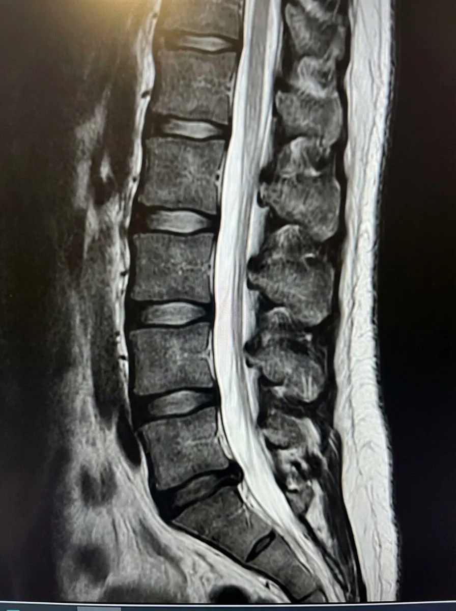 |
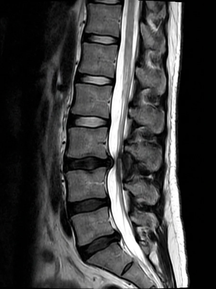 |
These are illustrative images, not actual MRI scans.