"Why Does It Get Narrow?" — How Spinal Stenosis Works
Inside your spine, there's a "tunnel" where your nerves travel.
Last week, we talked about how lumbar spinal stenosis affects an estimated 103 million people worldwide — it's far from a rare condition. This week, I'd like to explain why the spinal canal narrows in the first place — as simply and clearly as I can.
Understanding the Structure of Your Spine
Let me start with a quick look at how your spine is built.
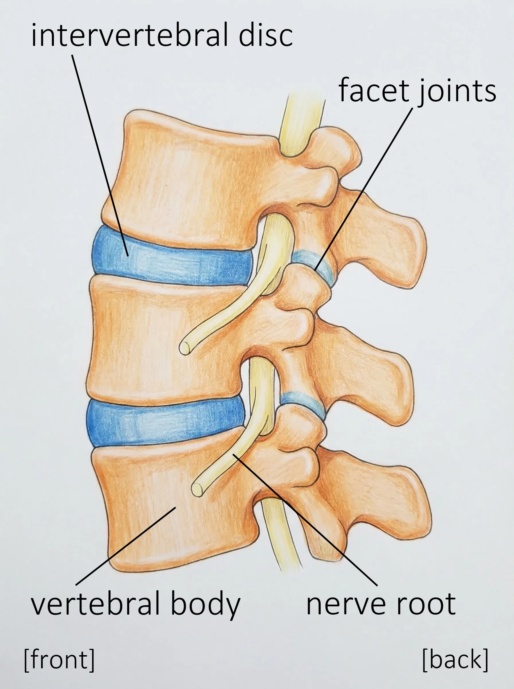
Your spine is made up of individual bones called vertebrae, stacked on top of each other like building blocks. In the lower back, there are five of these bones, and together they're called the lumbar spine.
Between each vertebra sits a disc — a cushion-like structure filled with water and gel that absorbs shock when you move.
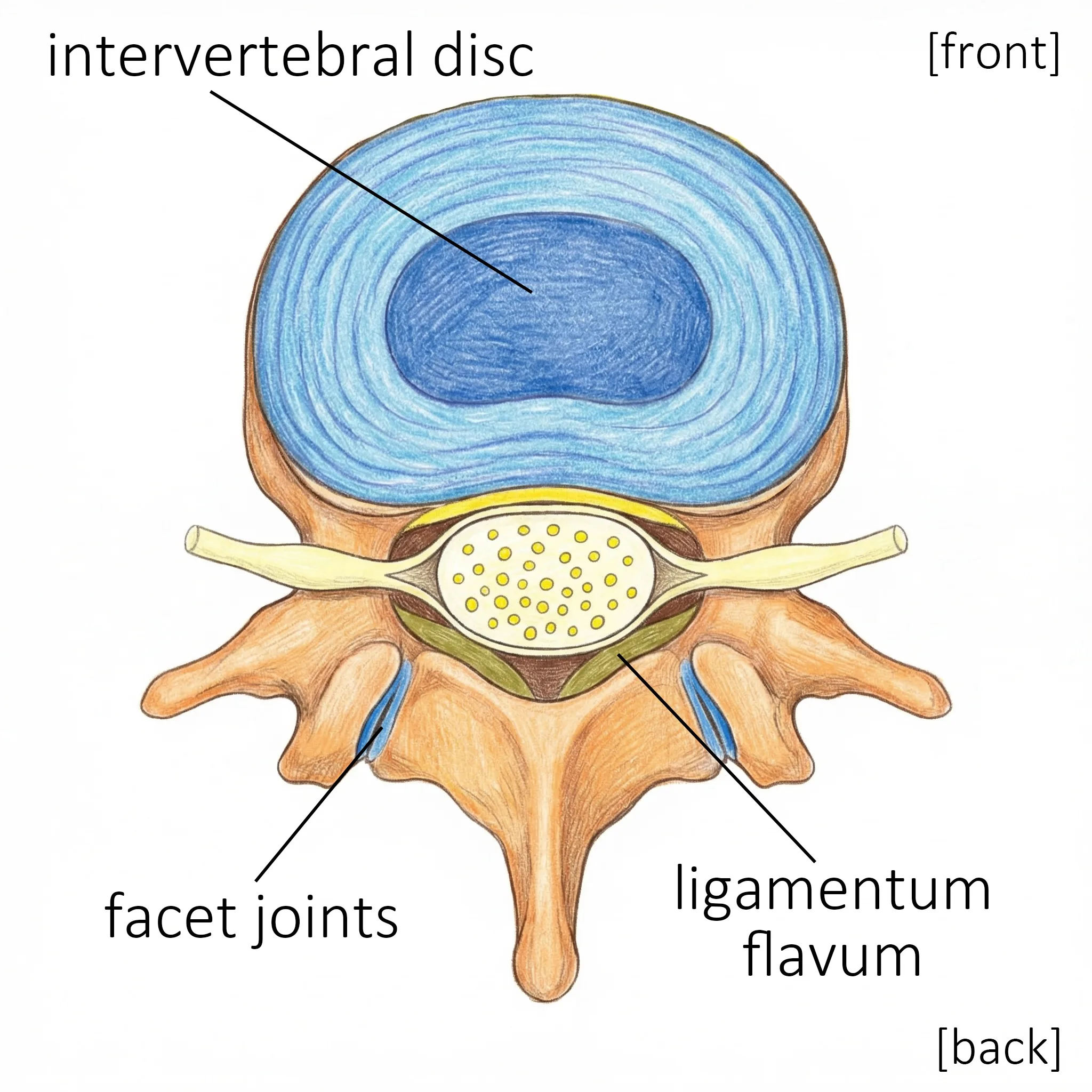
When you stack these vertebrae on top of one another, there's a hollow space that runs down the center. This is the spinal canal.
Inside this canal runs a bundle of nerves extending from the brain. In the lower back, these nerves are called the cauda equina (literally "horse's tail"), and they control sensation and movement in your legs, as well as bladder and bowel function.
Here's a helpful way to picture it:
Imagine building blocks (vertebrae) stacked to form a tunnel (the spinal canal), with a precious electrical cable (nerves) running through the middle.
Three Reasons the Tunnel Gets Narrower
As we age, the parts that make up this tunnel gradually wear down and change shape. This is the main cause of spinal stenosis.
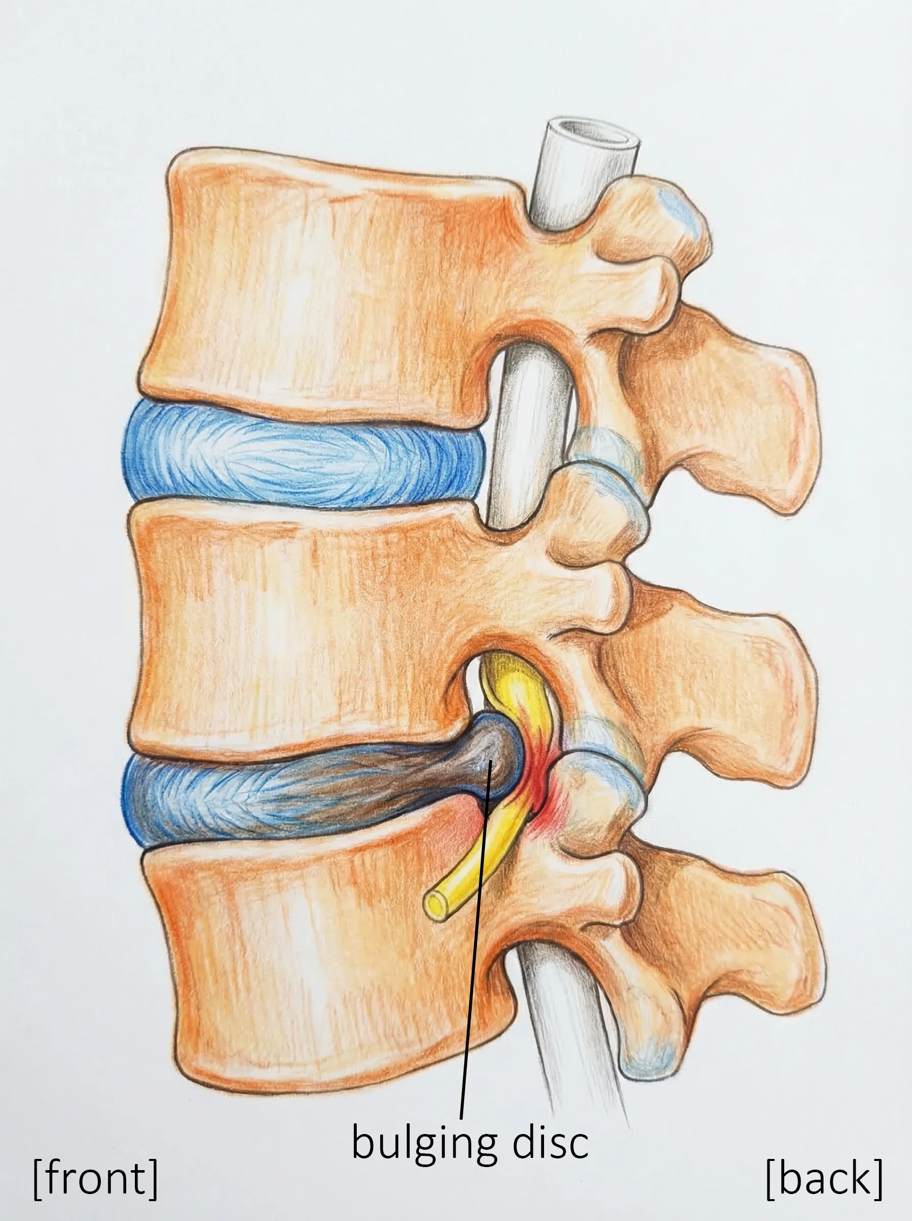
1. Bulging Discs
Over time, the discs between your vertebrae lose water content and elasticity. As they flatten out, they can bulge into the spinal canal, taking up space.
Think of it this way: A new cushion is plump and firm, but after years of use, it flattens and spreads outward. The same thing happens to your spinal discs.
2. Thickening of the Ligaments
The ligaments that hold your spine together — strong bands that connect bone to bone — can thicken with age. In particular, the ligamentum flavum (a yellow-colored ligament at the back of the spinal canal) can become much thicker than normal, squeezing the tunnel from behind.
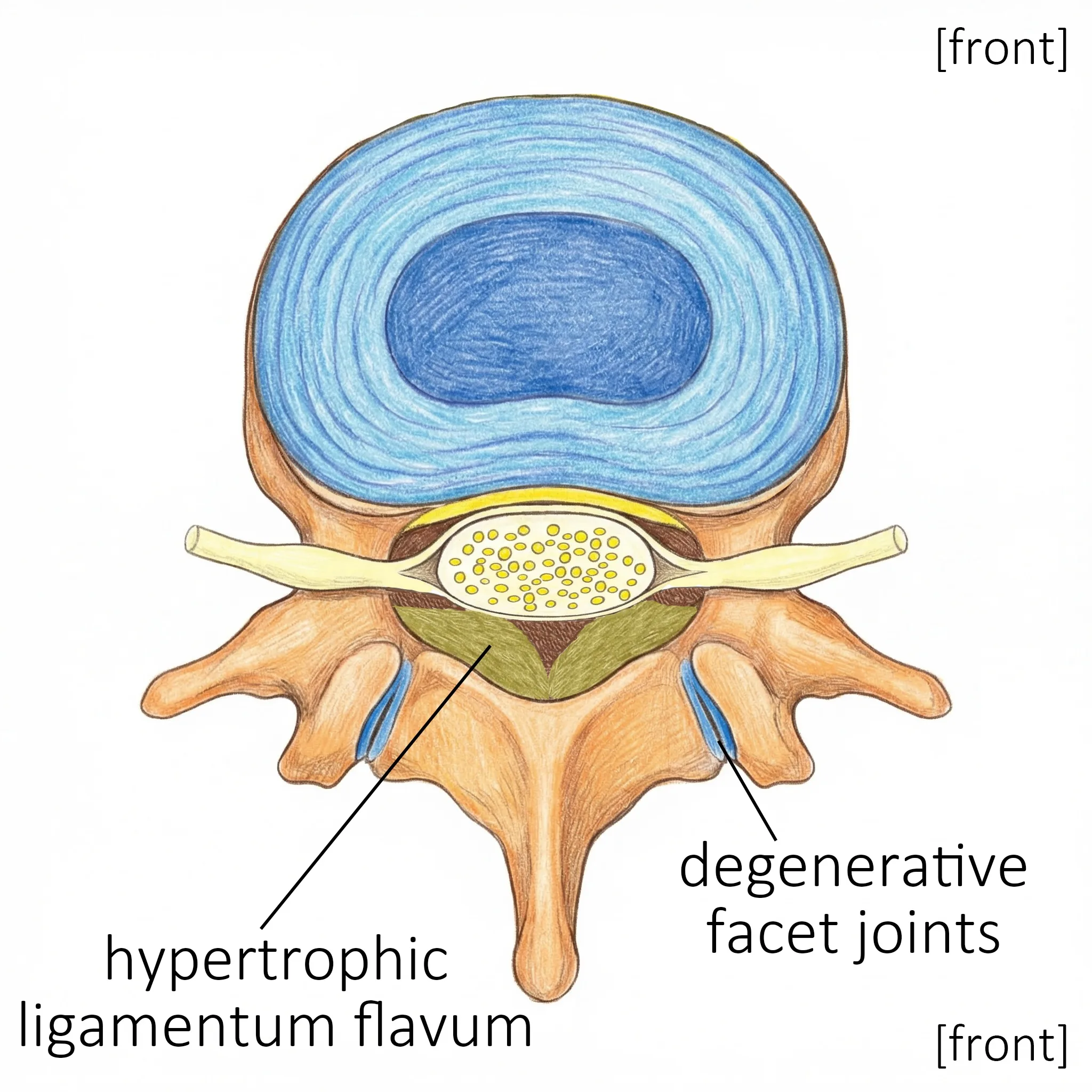
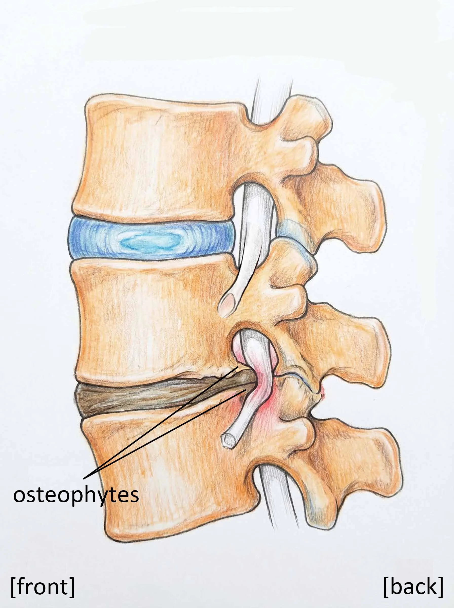
3. Bone Spurs (Osteophytes)
Small bony growths called osteophytes (bone spurs) can develop along the edges of the vertebrae. These are part of the aging process and cause the walls of the tunnel to protrude inward.
These three changes, often occurring together, cause the nerve tunnel to narrow gradually over time.
Here's something important to keep in mind: These changes don't happen overnight. They develop slowly, over many years. And not everyone who has narrowing will experience symptoms. Many people whose MRI scans show a narrow spinal canal have no pain or discomfort at all.
The Connection with Spondylolisthesis
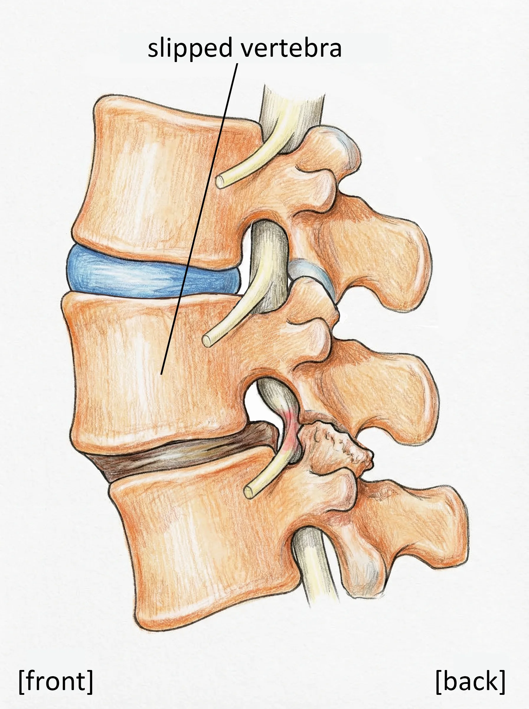
A condition you may hear about alongside spinal stenosis is spondylolisthesis — where one vertebra slips forward over the one below it.
When a vertebra slips out of position, it narrows the spinal canal on its own. Rather than being a completely separate condition, spondylolisthesis and stenosis frequently occur together.
If spondylolisthesis is present, it may affect the type of surgery recommended. (We'll cover this in detail in Week 9.)
What If Your MRI Shows "Stenosis"?
Here's something very important I'd like you to remember:
An MRI image alone does not determine whether surgery is needed.
Many people whose scans show narrowing have only mild symptoms. On the other hand, some people with relatively normal-looking scans experience significant pain and disability.
What matters most is a comprehensive evaluation that combines imaging findings with your actual symptoms. A finding of stenosis on MRI does not automatically mean you need surgery.
We'll talk more about diagnostic tests in Week 4.
It's Not Your Fault
I'd like to leave you with one more important thought.
Spinal stenosis is a result of completely natural changes that come with age. It doesn't happen because of bad posture, not exercising enough, or lifting heavy objects.
Of course, some people are born with a naturally narrow spinal canal (congenital stenosis). That's simply a variation in body type — and it's nobody's fault either.
These are changes that have occurred in a spine that has supported your body faithfully for decades. Rather than blaming yourself, the best approach is to understand the condition and respond appropriately.
Key Takeaways
- The spinal canal is a tunnel inside your spine through which nerves travel
- With age, discs, ligaments, and bones change, causing the tunnel to narrow
- These changes happen gradually, and not everyone develops symptoms
- MRI findings alone don't determine the course of treatment
- This is a natural part of aging — it is not your fault