What tests you'll need, what they reveal, and how your doctor reaches a diagnosis of lumbar spinal stenosis.
Knowing what each test shows can help you feel prepared.
What You'll Learn
What your doctor will ask during the consultation
What happens during the physical examination
Imaging tests (X-ray, MRI, CT)
How to understand your test results (overview)
Second opinions
1. How the Consultation Works
1. The Medical Interview
Your doctor will start with detailed questions about your symptoms.
Questions You May Be Asked
Question
Why It Is Asked
"When did the pain start?"
To determine whether this is acute or chronic
"Where does it hurt?"
To find out whether only the back is affected, or the legs as well
"When does it hurt most?"
To understand the relationship between posture, activity, and pain
"How far can you walk?"
To assess the severity of intermittent claudication
"Do you have any numbness?"
To check for nerve involvement
"Any problems with urination or bowel control?"
To assess severity (this may indicate an emergency)
"Have you had any previous back treatments?"
To review your treatment history
Information That Helps Your Doctor
Before your appointment, it may help to jot down:
Where the pain is: lower back, buttock, thigh, calf, foot, etc.
How far you can walk continuously: in meters or minutes
Positions that bring relief: leaning forward, sitting, etc.
How daily life is affected: shopping, housework, work, etc.
Medications you are currently taking
You may feel that your doctor did not ask enough about the detailed timeline of your pain. The exact order of events matters less than you might expect. Briefly say when the pain started and where it hurts.
2. The Physical Examination
After the interview, your doctor will examine you.
Key Tests
Test
How It Is Done
What It Reveals
Visual inspection
Observing your posture and how you walk
Deformity, abnormal gait
Palpation
Pressing on your lower back
Tender spots, muscle tension
Range of motion
Bending forward and backward
Which movements provoke pain
Neurological exam
Testing reflexes, sensation, and strength
Which nerve is affected
SLR test
Raising your leg while lying on your back
Helps distinguish a herniated disc
Neurological Examination in Detail
Test
How It Is Done
What It Means
Knee reflex (patellar reflex)
Tapping below the kneecap with a reflex hammer
Assesses the L4 nerve
Ankle reflex (Achilles reflex)
Tapping the Achilles tendon
Assesses the S1 nerve
Sensory testing
Checking your ability to feel touch
Identifies which nerve is affected
Muscle strength testing
Testing the power of your ankles and toes
Assesses nerve motor function
2. Imaging Tests
X-Ray
Purpose
An X-ray examines the condition of your bones.
What It Can and Cannot Show
What It Can Show
What It Cannot Show
Bone deformity (bone spurs)
The condition of your nerves
Disc height (indirectly)
Soft tissues (discs, ligaments)
Spondylolisthesis (slipping)
The degree of stenosis
Scoliosis, kyphosis
Herniated discs
Key Features
Quick and simple; it can be done right away
Uses a small, low dose of radiation
Taken while standing, so it shows how your spine looks under your body weight
Can show instability: by comparing images taken while you bend forward and lean back, your doctor can see whether a vertebra shifts with movement
MRI (Magnetic Resonance Imaging)
Purpose
MRI gives detailed images of your nerves and soft tissues. It is the most important test for diagnosing spinal stenosis.
What It Shows
What It Can Show
The degree of spinal canal narrowing
Whether nerves are being compressed
The condition of your discs
Thickening of the ligaments
Herniated discs
Features
Advantages
Disadvantages
Excellent detail of nerves
The scan takes time (20-40 minutes)
No radiation
You lie inside a narrow tube
Shows soft tissues clearly
The machine is loud
Discuss with your doctor if you have metal implants
Pacemakers and SCS devices require special precautions
Tips for Your MRI
Stay still. Movement blurs the images.
If small spaces make you anxious (claustrophobia), let your doctor know beforehand.
Take off jewelry, accessories, and other metal items.
Tattoos carry a small risk of skin burns; tell your doctor in advance.
CT Scan (Computed Tomography)
Purpose
A CT scan gives detailed images of your bones.
What It Shows
What It Can Show
Detailed bone anatomy
Location and size of bone spurs
The neural foramen (the opening where nerves exit)
Surgical planning information
Features
Advantages
Disadvantages
Excellent bone detail
Involves radiation
Quick scan time
Does not show nerves as well
More comfortable than MRI
Contrast dye may be used
Myelography
Before MRI became widely available, doctors injected a contrast dye into the spinal sac and then took X-rays and CT scans. This is called myelography. Because it is invasive, MRI has largely replaced it for diagnosis. Myelography may still be done when the doctor suspects that spinal movement (bending forward and backward) plays a major role in your symptoms, because a standard MRI is taken while you lie still.
How Tests Are Combined
Tests are usually done in this order:
[!note] Typical sequence of tests
X-ray: an overall picture of the bones
MRI: a detailed look at the nerves
CT (if needed): confirms bone details (especially if fusion surgery is being considered)
3. Your Results and Second Opinions
Understanding Your Test Results (Overview)
What Stenosis Looks Like on MRI
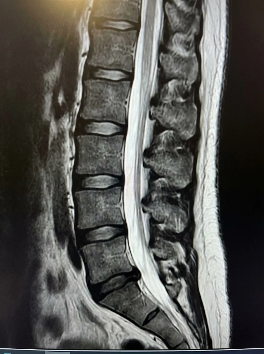
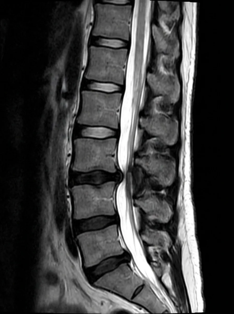
MRI can show how narrow the spinal canal has become.
The MRI images below compare a normal spinal canal with one that has stenosis. In a normal spine, the bright white cerebrospinal fluid fills the spinal canal with plenty of room. In stenosis, the canal is narrowed.
Normal
Stenosis
These are illustrative images, not actual MRI scans.
An Important Point
Narrowing on imaging does not equal severity of symptoms.
Some people have significant narrowing on MRI but no symptoms. Others have only mild narrowing but severe symptoms.
Imaging alone does not determine your treatment plan. Your doctor considers both your symptoms and your imaging findings together.
Second Opinions
What Is a Second Opinion?
Asking another doctor for their view is called getting a second opinion.
When to Consider One
Situation
Example
You are not convinced about the diagnosis
"Is this really what I have?"
You are unsure about the treatment plan
"Should I have surgery?"
Before making a major decision
"I want to check before going ahead with surgery"
You want to know about other options
"Are there other treatment approaches?"
The Second Opinion Process
[!note] The second opinion process
Discuss with your current doctor
Ask for a referral letter and your test results
Book a second opinion appointment
Hear the other doctor's assessment
Make your own decision about treatment
Things to Keep in Mind
Tell your current doctor. This is not rude; it is your right as a patient.
Bring your test data. You will need your MRI images (usually on a disc or USB).
Check the cost. Insurance may not always cover a second opinion consultation.
4. Common Questions, Summary, and Questions for Your Doctor
Frequently Asked Questions
Q. Is an MRI painful?
A. It does not hurt. You do lie inside a narrow tube, which can feel uncomfortable if you have claustrophobia. Let your doctor know in advance if this concerns you.
Q. How soon will I get my results?
A. X-ray results are usually available the same day. MRI results may be explained at a follow-up visit, because a specialist needs to review the images carefully. In many healthcare systems, an MRI cannot be performed on the same day as your first consultation unless emergency surgery is being considered.
Q. Do I need a contrast MRI?
A. In most cases, spinal stenosis can be diagnosed without contrast dye. Contrast may be used if a tumor is suspected or if a more detailed evaluation is needed.
Q. How much do these tests cost?
A. Costs vary depending on your country and insurance coverage. In the United States, with insurance, you can generally expect:
X-ray: $50-150
MRI: $250-500
CT: $150-300
Check with your provider for specific out-of-pocket costs.
Summary
Key Point
Details
Medical interview
Describe your symptoms clearly
Physical examination
Assesses the state of your nerves
X-ray
Shows the overall bone structure
MRI
The most important test for diagnosing stenosis
CT scan
Shows bone in detail
The key principle
Treatment decisions are based on both symptoms and imaging, not imaging alone
Consider asking these questions during your consultation or after receiving test results:
"Is the stenosis on my MRI mild, moderate, or severe?"
This gives you an objective measure of the narrowing
"Do my symptoms match the findings on my imaging?"
This tells you whether the imaging findings explain your symptoms and whether the treatment plan makes sense
"At which level is the stenosis most severe (e.g., L3/4, L4/5)?"
This identifies the main problem area
"If there is stenosis at multiple levels, which one is the main source of my symptoms?"
In multi-level stenosis, this clarifies the treatment target
"Based on my test results, how quickly might this progress?"
This tells you whether close monitoring or early treatment is needed
"Do I need any additional tests (contrast MRI, CT myelography, etc.)?"
This confirms whether the current diagnosis is complete or more testing is needed
"Is the nerve compression reversible? How much recovery can I expect from treatment?"
This helps you set realistic expectations
"If I want a second opinion, what records should I bring?"
Typically you will need a referral letter, MRI images, and blood test results
Tip:
When your doctor explains your test results, refer to the "What Stenosis Looks Like on MRI" diagram on this page and ask, "Which of these pictures is closest to mine?" The explanation becomes much easier to follow.
This page provides medically accurate information, but it is not a substitute for professional medical advice. Please consult your doctor for diagnosis and treatment tailored to your individual situation.