To understand your condition, it helps to start with how your spine is built.
What You'll Learn
- How the spine is structured
- What the "spinal canal" is
- Where your nerves travel
- Why the spine changes with age
A clear guide to spinal anatomy — the vertebrae, nerves, and structures that matter when understanding spinal stenosis.
To understand your condition, it helps to start with how your spine is built.
What You'll Learn
- How the spine is structured
- What the "spinal canal" is
- Where your nerves travel
- Why the spine changes with age
Your spine is made up of individual bones called vertebrae, stacked on top of one another like a tower of building blocks.

In total, your spine has 24 vertebrae plus the sacrum at the bottom.
When we talk about lower back pain and spinal stenosis, we usually mean the lumbar spine, the five vertebrae in your lower back.
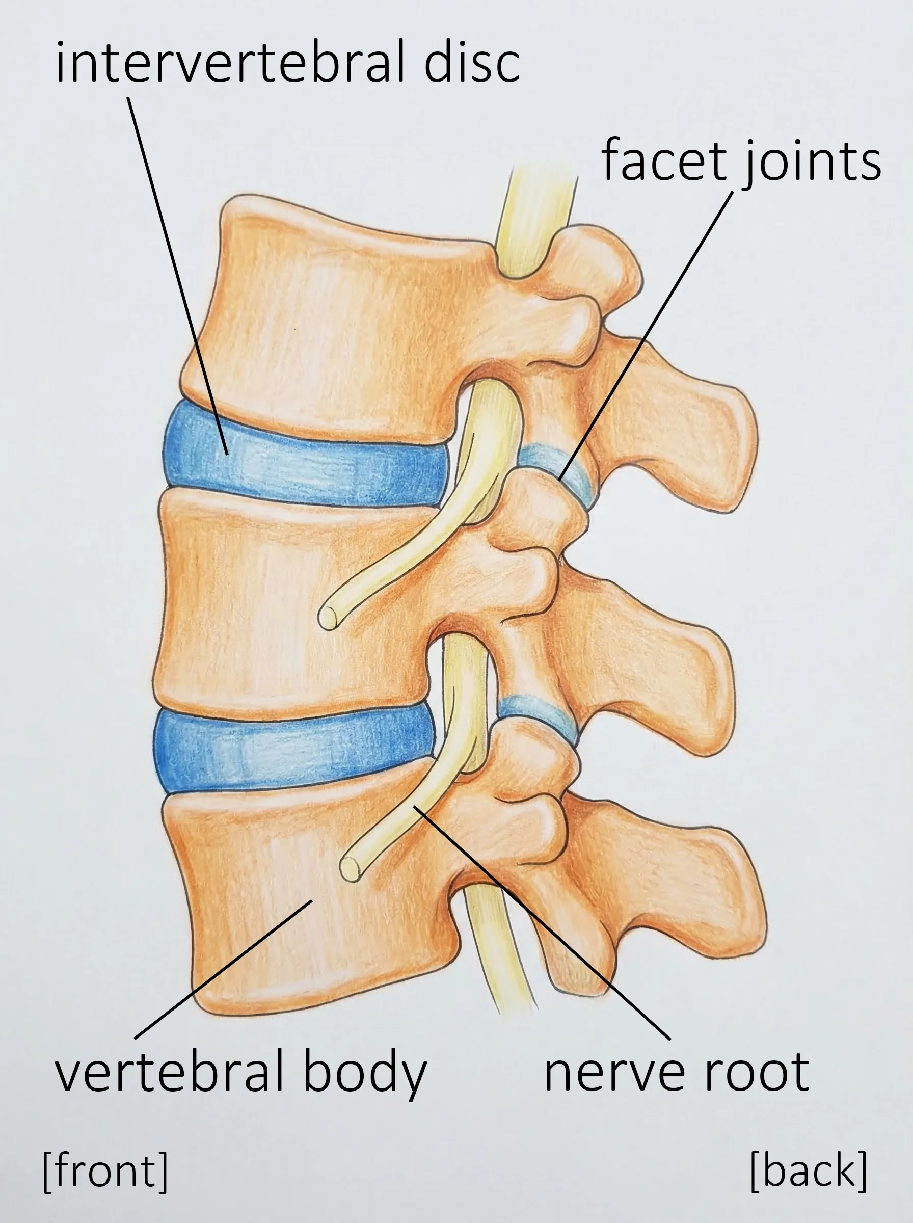
Each vertebra has a hole in its center. When the vertebrae are stacked together, these holes line up to form a continuous tunnel running down the length of your spine. This tunnel is called the spinal canal.
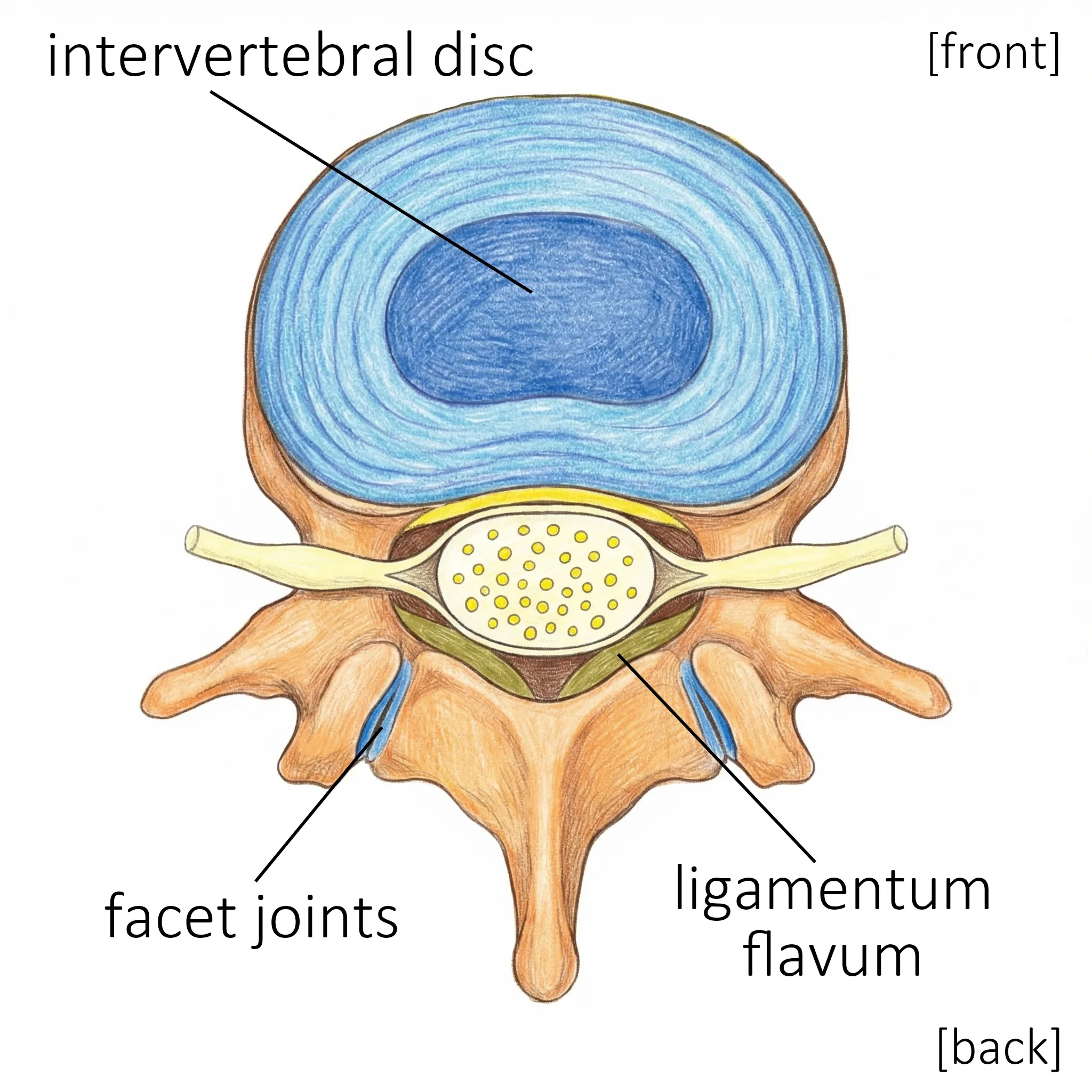
Think of it this way: The spinal canal is like a garden hose about 2–3 centimeters (roughly 1 inch) in diameter. Your nerves travel inside this hose.
There are three types of nerve structures to know about:
The spinal cord is a thick bundle of nerves that carries signals between your brain and your body, like a communications cable. It relays your brain's commands to your muscles and sends sensations (touch, temperature, pain) back to your brain.
The spinal cord belongs to the central nervous system and typically ends at the level of the 1st or 2nd lumbar vertebra.
Important: About Spinal Cord Injuries Because the spinal cord is part of the central nervous system, like the brain, damage to it is often permanent and irreversible. As with the lasting effects of a stroke, there is currently no way to fully repair spinal cord injuries.
Below where the spinal cord ends (below the 2nd lumbar vertebra), the spinal cord fans out into a bundle of individual nerve strands. These strands resemble a horse's tail, which is why they are called the cauda equina (Latin for "horse's tail").
The cauda equina belongs to the peripheral nervous system. So the chance of recovery is better than with spinal cord injuries, although severe damage can still lead to permanent problems.
At each level of the spine, a pair of nerve roots branches off, one to the left and one to the right, and exits the spinal canal.
These nerve roots also belong to the peripheral nervous system. The nerve roots from the cervical spine control your arms and hands; those from the thoracic spine become the intercostal nerves (between your ribs); and those from the lumbar spine control your legs and feet.
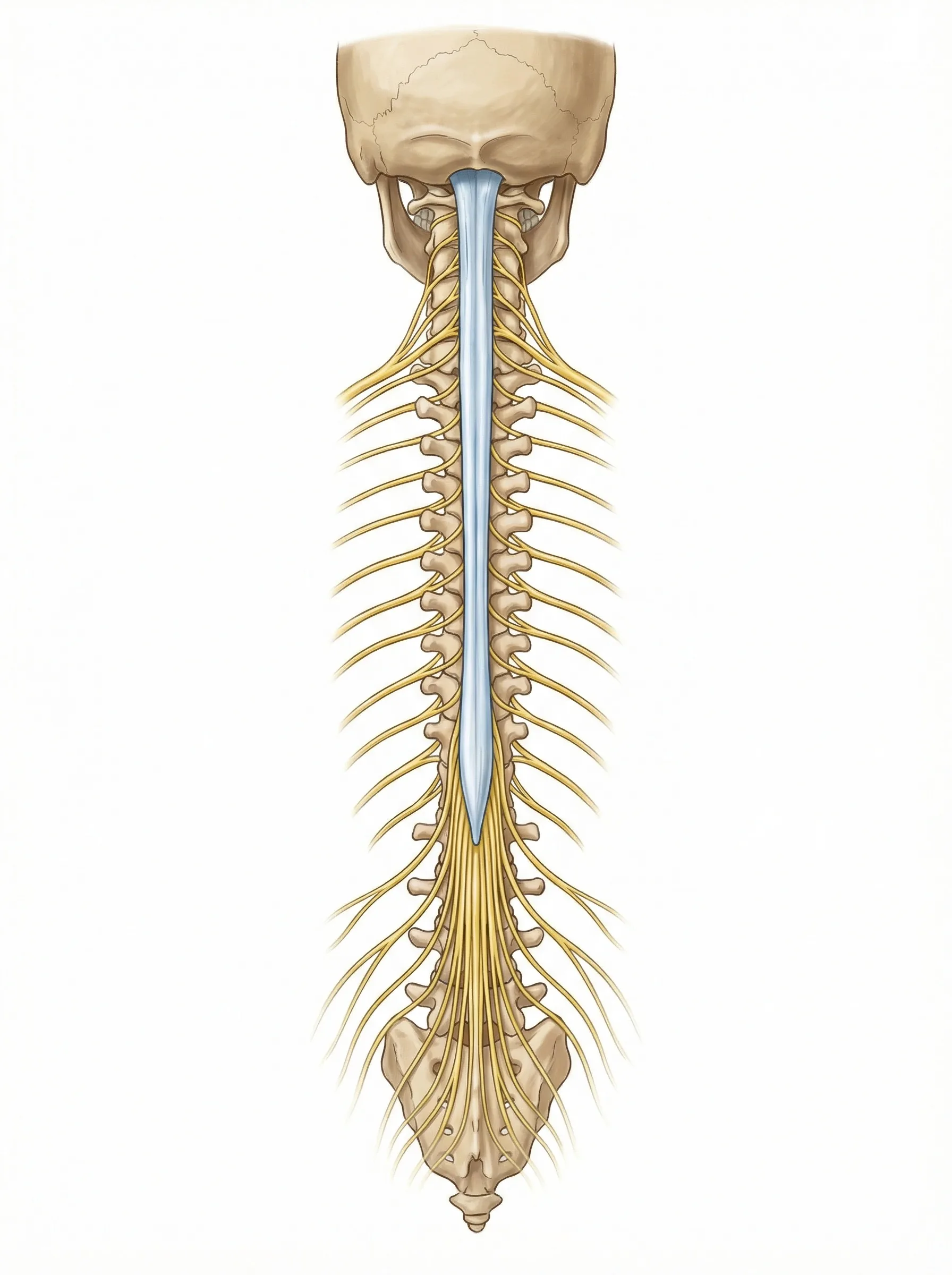
Because the cauda equina and nerve roots are peripheral nerves, they have some ability to recover, unlike the spinal cord.
However, if both legs become paralyzed, this is a medical emergency.
Go to the hospital immediately if you experience:
If these symptoms develop, permanent paralysis can result unless you receive treatment quickly.
Do not wait and see. Go to the emergency room right away.
Behind the spinal canal, there are arch-shaped bones called the laminae that form a protective roof over the nerves. On either side of the laminae are the facet joints.
These joints connect the vertebrae above and below, and they guide and support the movement of your spine.
When your spine bends:
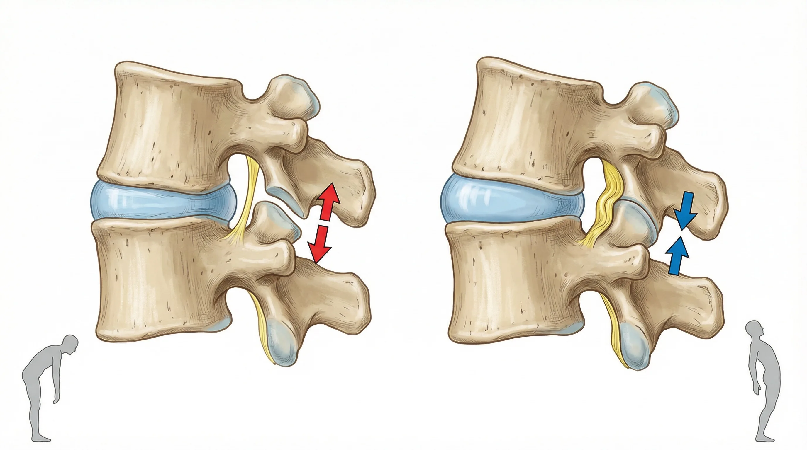
Think of it this way: Facet joints have a structure similar to your knee joint.
Decades of bending and straightening can lead to knee arthritis. In the same way, your facet joints change after decades of repeated movement:
This thickening is one of the reasons the spinal canal becomes narrower.
The vertebrae are connected to one another by strong bands of tissue called ligaments.
In spinal stenosis, two ligaments matter most:
This is the single most important ligament in lumbar spinal stenosis.
The ligamentum flavum lines the back wall of the spinal canal from the inside.
This ligament connects the spinous processes, the bony bumps you can feel along your back. Like the ligamentum flavum, it helps control how far the spine can bend forward.
Here is what the ligaments do with each movement:
When you bend forward:
When you lean backward:
After decades of being stretched and released over and over:
→ The ligamentum flavum gradually thickens
→ The thickened ligament compresses the spinal canal from behind
→ This is one of the major causes of spinal stenosis
In simple terms: A rubber band that is stretched and released for years also becomes thicker and stiffer.
When the nerve compression is significant, how the ligamentum flavum behaves during movement strongly affects your symptoms. When you lean backward, the ligament bunches up and presses on the nerves, making leg pain worse. When you lean forward, the ligament is stretched out and pressure on the nerves is relieved. This is why leaning forward eases your symptoms and leaning backward makes them worse. It is a typical pattern of lumbar spinal stenosis, and the ligamentum flavum drives it.
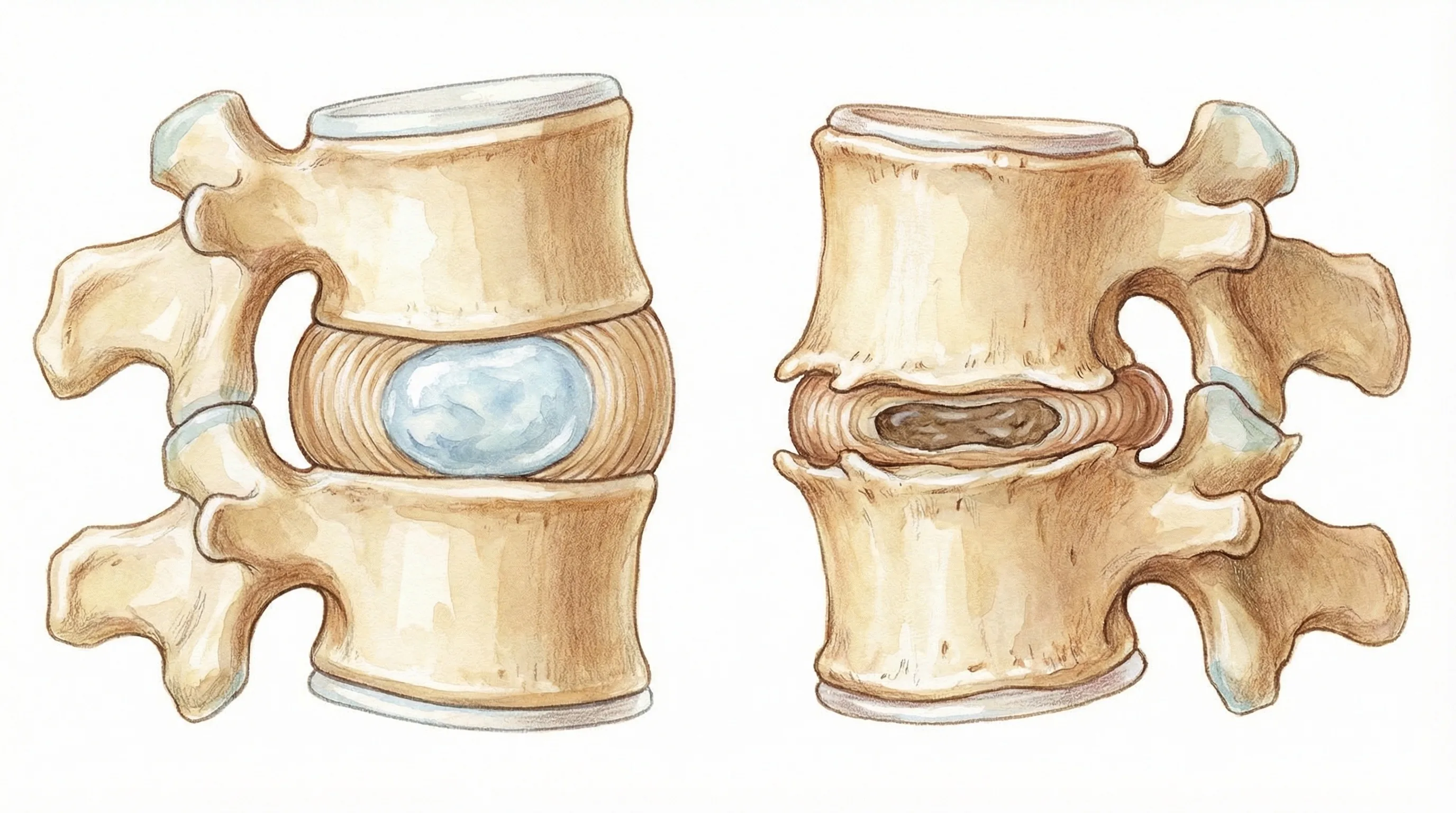
Between each pair of vertebrae sits an intervertebral disc, a cushion made of cartilage.

Each disc has a nucleus pulposus, a jelly-like center, surrounded by the annulus fibrosus, a tough outer ring of fibers.
Think of it this way: A disc is like a jelly doughnut:
These discs let your spine bend in all directions and absorb shocks from walking, running, and daily activities.
Spinal aging begins in your 20s.
A little-known fact: Disc degeneration starts earlier than most people expect. Because it progresses so slowly, symptoms often don't appear until decades later.
The nucleus pulposus (the jelly-like center of the disc) gradually loses water content over the years.
→ As it dries out, the disc loses its ability to support weight effectively
As the nucleus shrinks, the overall thickness of the disc decreases.
A familiar example: You may have noticed that you're a bit shorter than you were when you were younger. A major reason is disc degeneration. With 23 discs in your spine, even a loss of just 1–2 millimeters per disc adds up to a height reduction of 2–4 centimeters (about 1–1.5 inches).
As the disc loses height, the remaining annulus fibrosus (the tough outer ring) has nowhere to go and begins to
→ Bulge beyond the edges of the vertebral body
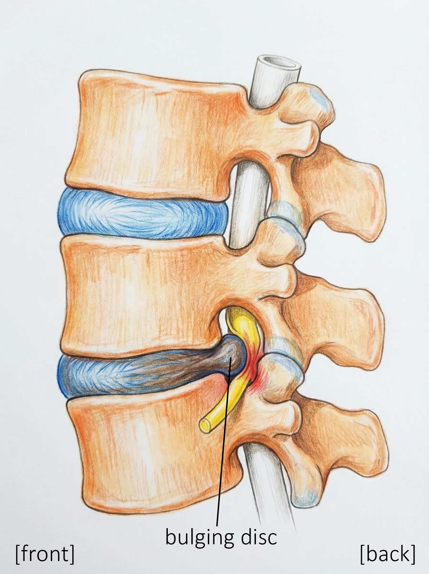
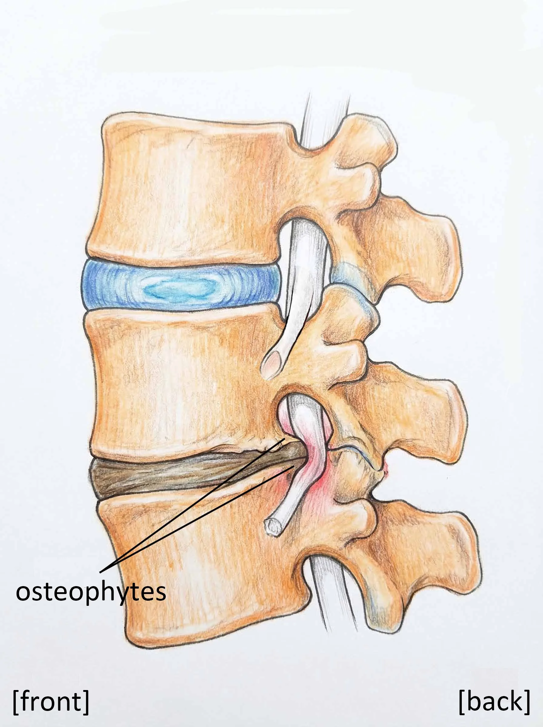
When the bulging annulus irritates the edges of the vertebral body:
→ The bone responds by growing outward
→ These bony outgrowths are called osteophytes (bone spurs)
Osteophytes can contribute to nerve compression.
| Region | Range of Motion | Age-Related Changes |
|---|---|---|
| Cervical (neck) | Moves a great deal | Prone to degeneration |
| Thoracic (mid-back) | Limited movement (attached to ribs) | Less affected |
| Lumbar (lower back) | Bears heavy loads and moves a great deal | Most affected |
The general rule: the more a section of the spine moves, the more susceptible it is to age-related wear.
That is why problems most commonly occur in the cervical spine (neck) and the lumbar spine (lower back).
Over time, several changes happen at the same time:
| Structure | When Young | With Aging |
|---|---|---|
| Discs | Plump and springy | Dried out and thinner |
| Bone (vertebral body) | Smooth edges | Osteophytes (bone spurs) form |
| Facet joints | Smooth and gliding | Misshapen and thickened |
| Ligaments (ligamentum flavum) | Thin and flexible | Thickened and stiff |
| Spinal canal | Spacious | Narrowed ← This is "stenosis" |
When these changes add up, the spinal canal becomes too narrow and the nerves inside are compressed. This is lumbar spinal stenosis. When the same degenerative process occurs in the cervical spine, it compresses the spinal cord in the neck and leads to a condition called cervical spondylotic myelopathy.
| Key Point | Details |
|---|---|
| Spine structure | 24 vertebrae stacked on top of one another |
| Spinal canal | A tunnel running through the vertebrae (like a garden hose) |
| Nerves | The spinal cord and nerve roots travel through the spinal canal |
| Discs | Cushions between the vertebrae (like a jelly doughnut) |
| Aging changes | The spinal canal narrows and may compress the nerves |
→ What Is Lumbar Spinal Stenosis?: the symptoms and causes of stenosis
→ Understanding Your Pain: why pain occurs and how it works
→ Tests and Diagnosis: how spinal stenosis is diagnosed
Consider asking these questions at your next appointment:
"Which part of my lumbar spine is affected?"
"How much have my discs degenerated?"
"At which level is the stenosis located?"
"Can you show me on my MRI where the nerves are being compressed?"
Tip: Feel free to bring the diagrams and tables on this page to your appointment. Pointing to an area and asking "Can you tell me more about this part?" is an easy way to start the conversation.
This page provides medically accurate information, but it is not a substitute for professional medical advice. Please consult your doctor for diagnosis and treatment tailored to your individual situation.