What lumbar spinal stenosis is, why it happens, and what symptoms to watch for — explained in plain language.
The medical name sounds complicated, but what is happening in your body is easy to understand.
What You'll Learn
What "lumbar spinal stenosis" means
Why this condition develops
How a normal spinal canal differs from a narrowed one
The types of stenosis (by structure and by symptoms)
An important message for you:
Do you ever think, "This pain isn't bad enough to mention"? Keeping quiet about pain makes it harder for your doctor to help you. Describing your pain accurately is the first step toward the right treatment. It's okay to say, "This hurts."
1. What Stenosis Is and Why It Happens
What Does "Lumbar Spinal Stenosis" Mean?
The term is easier to understand when you take it apart:
Term
Meaning
Lumbar
The lower back (the five vertebrae between your ribcage and pelvis)
Spinal canal
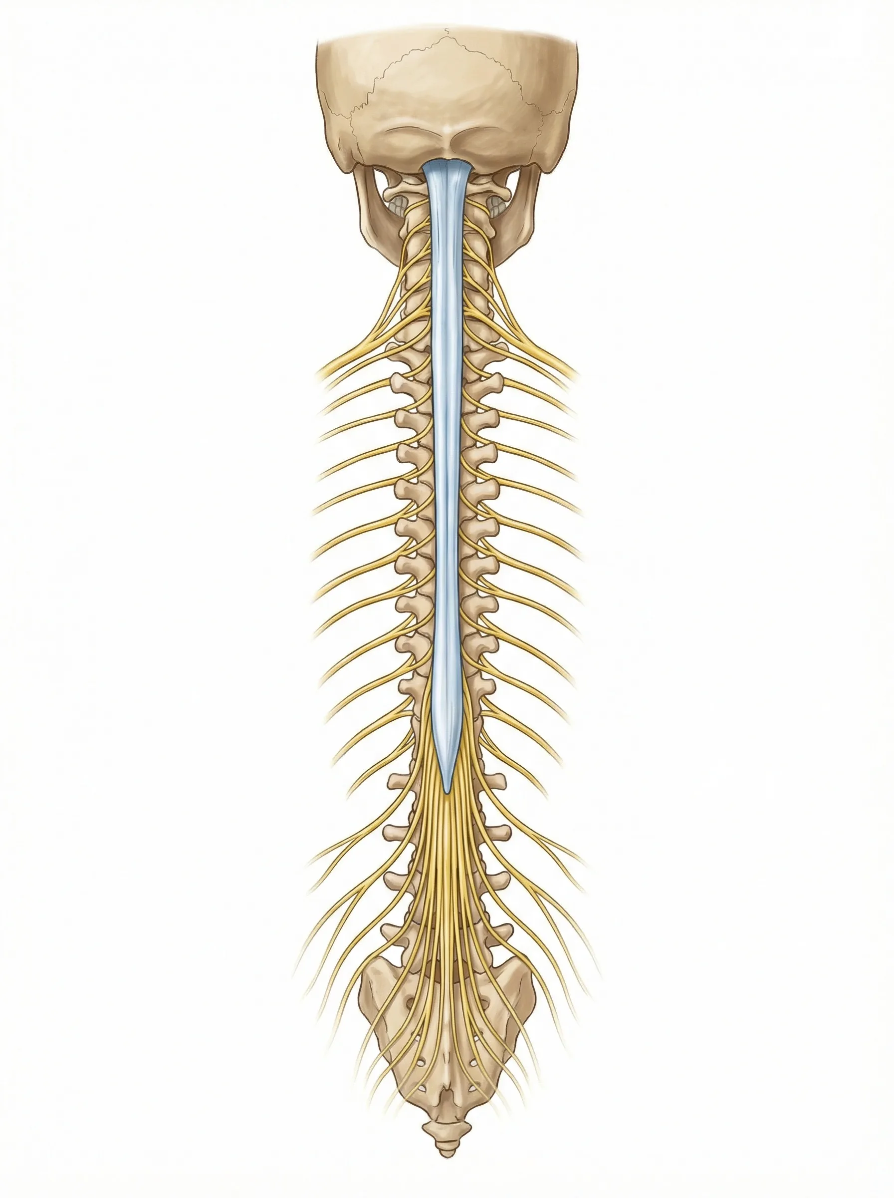
The tunnel inside your spine where the nerves travel
Stenosis
Narrowing
Put together, it means:
"A condition where the nerve tunnel in your lower back has become too narrow."
What's Happening Inside Your Spine
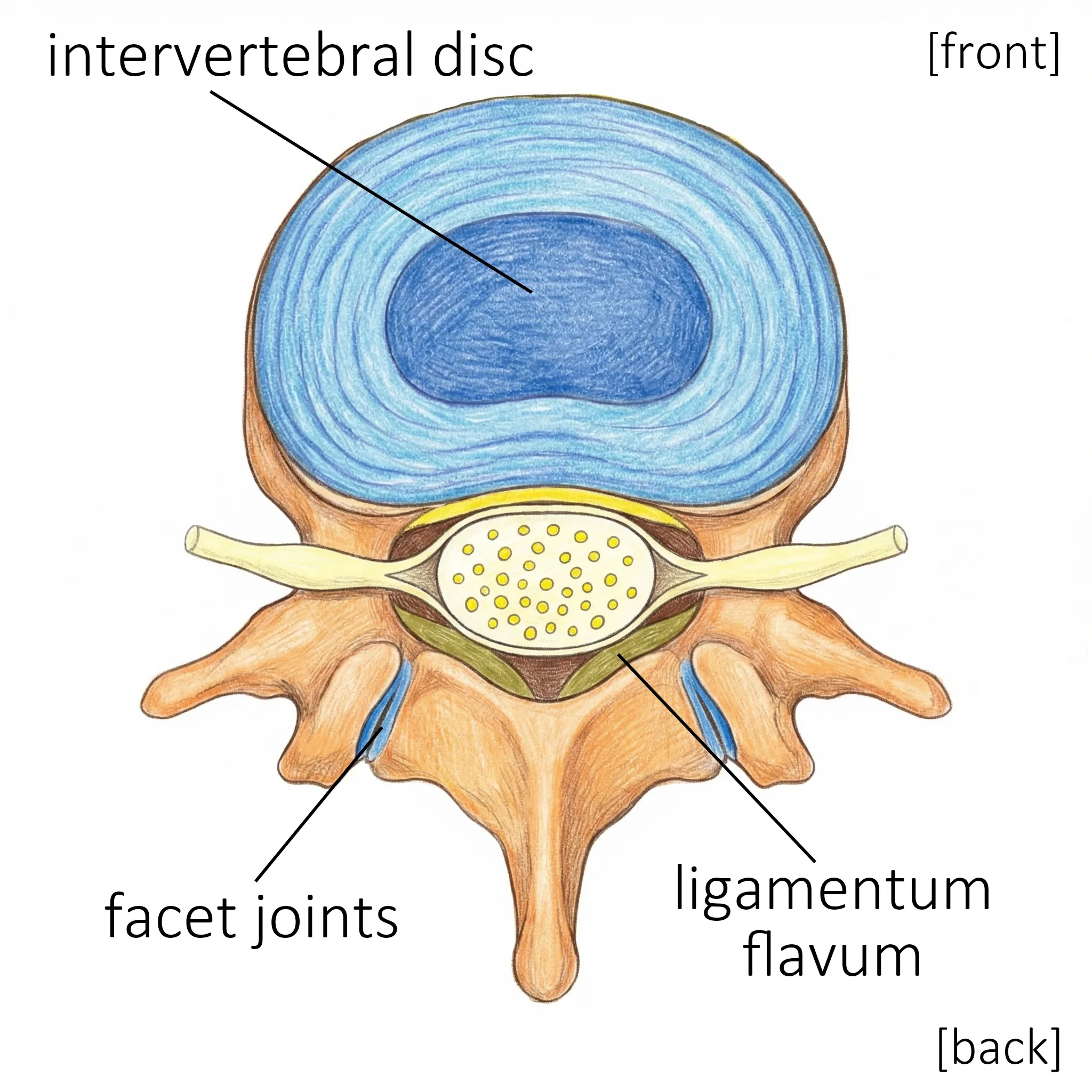
A Healthy Spine
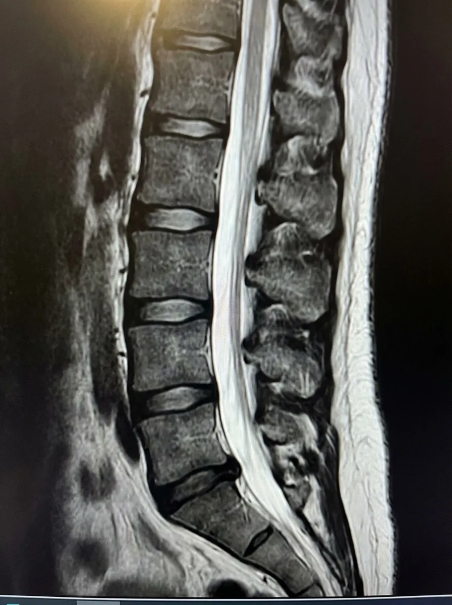
In a healthy spine, the spinal canal (the tunnel that protects your nerves) is wide enough for the nerves to pass through with room to spare.
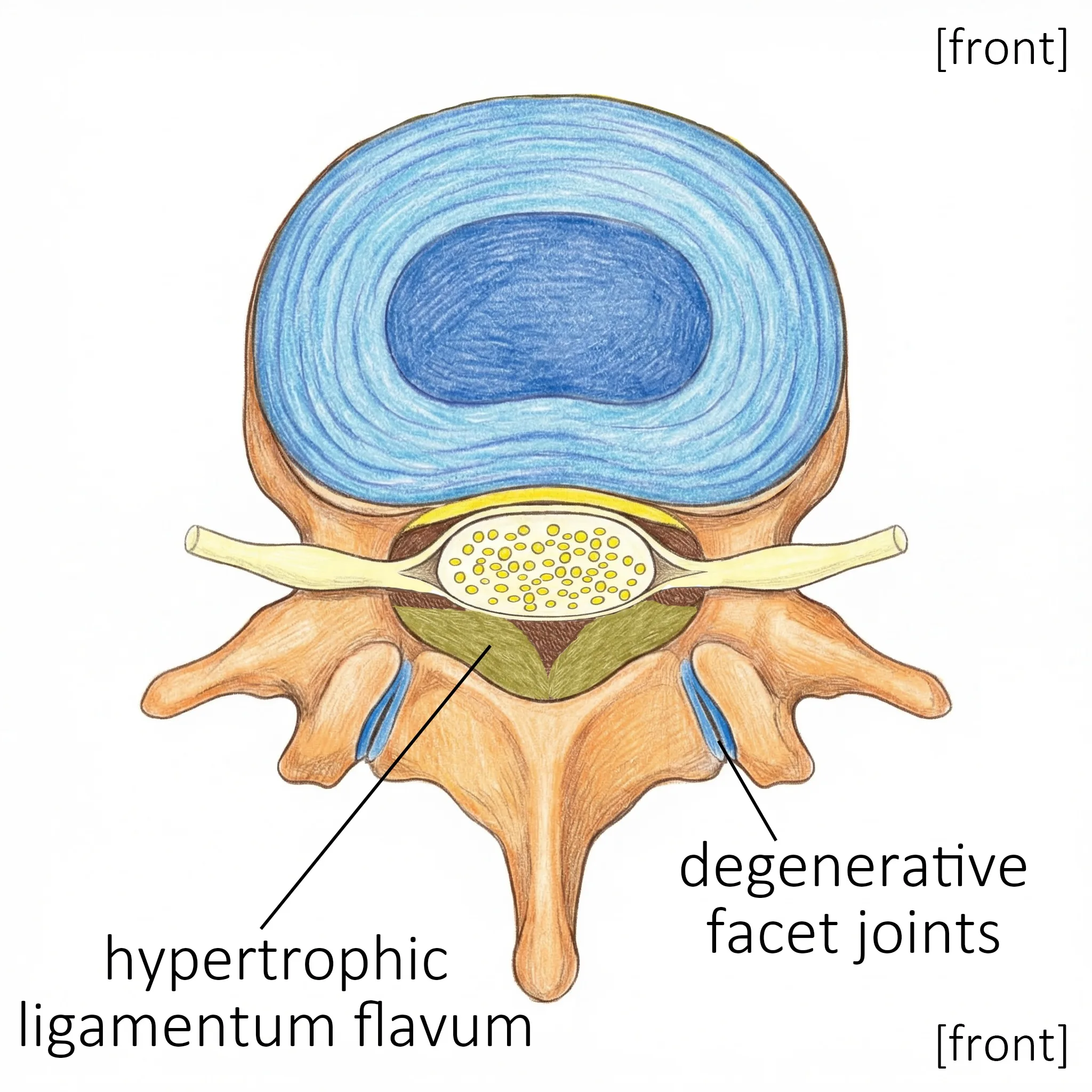
A Narrowed Spine
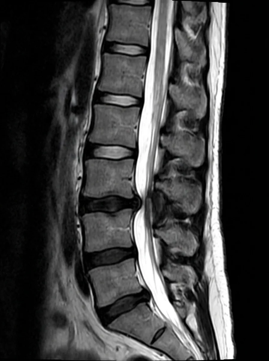
With spinal stenosis, the canal has become narrower, and the nerves are squeezed.
This pressure on the nerves causes pain, numbness, and tingling in your legs.
Why Does the Canal Become Narrow?
The spinal canal can narrow over time for several reasons.
Main Causes
Cause
Explanation
Age-related changes
The most common cause; a natural part of aging
Disc degeneration
The cushions between vertebrae wear down and may bulge into the canal
Bone spurs
Bony growths develop along the edges of the vertebrae
Thickened ligaments
The ligaments that support the spine grow thicker (especially the ligamentum flavum)
Degenerative spondylolisthesis
One vertebra slips forward over the one below it, narrowing the canal
Aging Is the Most Common Cause
Spinal stenosis is most common in people over 50 years old.
The everyday stresses on your spine cause small changes that add up over many years.
Something important to remember:
Spinal stenosis is not your fault. Nothing you did caused it. Everyone's spine changes with age, and in some people those changes are enough to press on the nerves. It is part of the body's natural aging, like gray hair or needing reading glasses.
Intermittent Claudication — The Hallmark Symptom
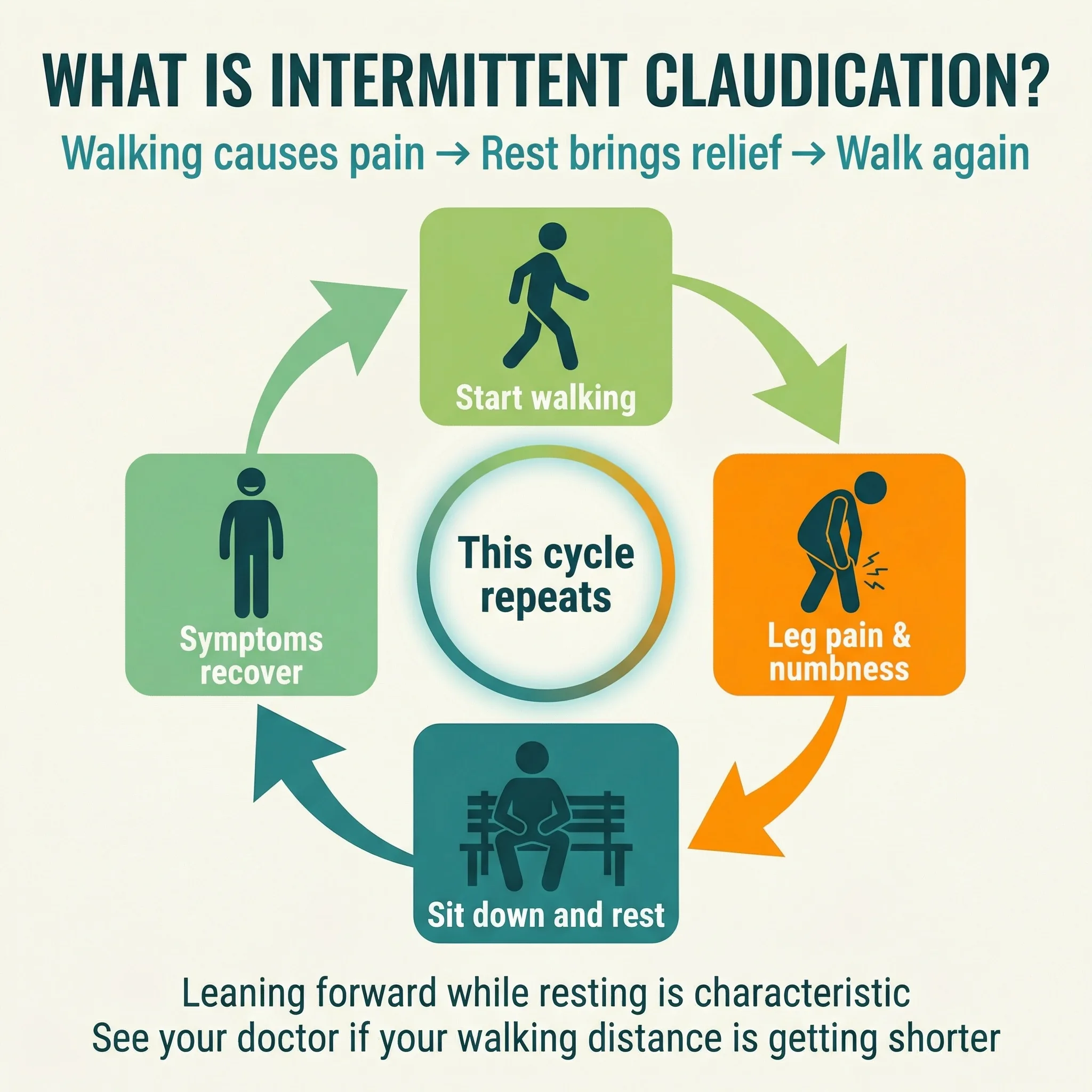
The most typical symptom of spinal stenosis is pain, numbness, or weakness in the legs that comes on during walking and goes away with rest. Doctors call this "neurogenic intermittent claudication." Leaning forward or sitting down usually brings relief. That is why many people with this condition can ride a bicycle without trouble even when walking is hard. If the distance you can walk comfortably is getting shorter, talk to your doctor.
2. Types of Stenosis
Types of Stenosis (by Location)
Stenosis is classified by where the narrowing occurs in the spinal canal.
1. Central Stenosis
The center of the spinal canal is narrowed.
What this means for you:
Symptoms tend to affect both legs
Walking may cause heaviness, tiredness, or numbness in both legs
2. Lateral Stenosis (Foraminal Stenosis)
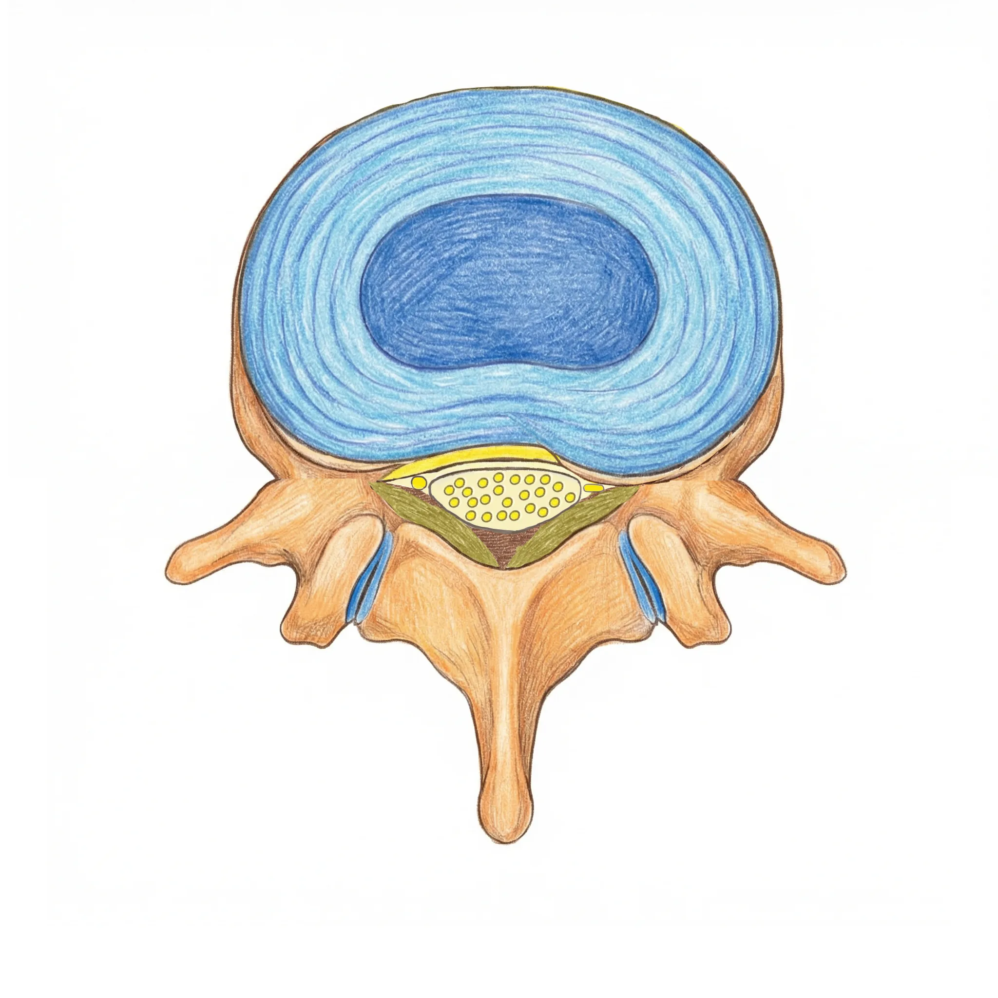
The nerve exit (called the foramen) on one side is narrowed.
A bony spur (osteophyte) at the edge of the vertebra presses on the nerve root on one side, close to its exit.
What this means for you:
Symptoms tend to affect one leg
Certain movements may make the pain worse
3. Combined (Mixed) Type
Both central and lateral narrowing are present. In practice, this combined type is the most common.
Types of Stenosis (by Symptoms) — Cauda Equina vs. Nerve Root
Doctors also classify stenosis by which nerves are affected and what symptoms result.
Type
What's compressed
Main symptoms
Cauda equina type
The bundle of nerves in the center of the canal (cauda equina)
Numbness in both legs, difficulty walking, bladder or bowel problems
Nerve root type
A single nerve branch (nerve root)
Pain from buttock down one leg
Mixed type
Both cauda equina and nerve root
Symptoms of both types
How the Two Classification Systems Relate
Structural type
Tends to cause...
Central stenosis
Cauda equina type symptoms (the central nerve bundle is compressed)
Lateral stenosis
Nerve root type symptoms (a nerve is pinched at its exit point)
Combined (structural)
Mixed type symptoms
Why does this matter to you?
Whether your stenosis is the cauda equina type or the nerve root type affects which medications work best. For example, the cauda equina type may respond well to medications that improve blood flow to the nerves (such as limaprost). The nerve root type is often treated with anti-inflammatory pain medications (NSAIDs). Ask your doctor: "Is my stenosis the cauda equina type or the nerve root type?"
3. Where It Occurs and How It Looks on Imaging
Where Stenosis Occurs Most Often
Stenosis can happen at any level of the lumbar spine, but some levels are affected more often:
Level
Location
How common
L4/5
Between the 4th and 5th lumbar vertebrae
Most common
L3/4
Between the 3rd and 4th lumbar vertebrae
Common
L5/S1
Between the 5th lumbar vertebra and the sacrum
Less common
Stenosis often occurs at more than one level (multi-level stenosis).
What It Looks Like on Imaging
X-rays can show some of the bony changes of stenosis, such as narrowed disc spaces, bone spurs, and vertebral slippage (spondylolisthesis). X-rays taken while you bend forward and lean back (flexion and extension X-rays) show your doctor whether there is instability, meaning the vertebrae shift more than they should when you move. X-rays cannot show the nerves themselves, so MRI (Magnetic Resonance Imaging) is needed for a complete picture.
On MRI, your doctor can see the spinal canal and the nerves directly, and confirm whether stenosis is present and how severe it is.
Normal
Stenosis
These are illustrative images, not actual MRI scans.
An Important Point
What the Images Show Does Not Always Match How You Feel
Some people have significant narrowing on MRI but feel no symptoms at all.Others have only mild narrowing but experience severe pain.
So treatment decisions are never based on images alone. Your doctor considers your symptoms together with the imaging findings to choose the right approach.
An MRI that shows stenosis does not by itself mean you need surgery. Doctors often list every change they see on an MRI. What matters is which of those changes are causing your symptoms.
4. Summary and Questions for Your Doctor
Summary
Key Point
Details
What it means
The nerve tunnel in your lower back has become too narrow
Main cause
Age-related changes in the discs, bones, and ligaments
Structural types
Central, lateral, or combined
Symptom types
Cauda equina, nerve root, or mixed; this affects medication choices
Most common level
L4/5 (between the 4th and 5th lumbar vertebrae)
Key takeaway
Imaging findings don't always match symptom severity
When you see your doctor, consider asking these questions:
"What type of stenosis do I have? Is it central, lateral, or combined? Is it the cauda equina type or nerve root type?"
Knowing both the structural type and the symptom type helps you make sense of your treatment options
"Is my stenosis mild, moderate, or severe?"
This tells you how advanced the condition is and what to expect
"Do my symptoms match what the MRI shows?"
This confirms whether the narrowing seen on imaging is causing your symptoms
"Is my stenosis likely to get worse over time?"
This tells you whether monitoring or preventive measures may be needed
"What treatment do you recommend at this stage?"
This starts a conversation about the options that suit your situation now
Tip:
Ask your doctor to show you your MRI images while explaining your condition. Seeing the pictures makes it much easier to understand. Many clinics can give you a copy of your images, so feel free to ask.
This page provides medically accurate information authored by an orthopedic spine surgeon. For diagnosis and treatment of your individual condition, please consult your physician.